« Previous card | Epithelial main | Next card »
Use the diagram above to complete the following:
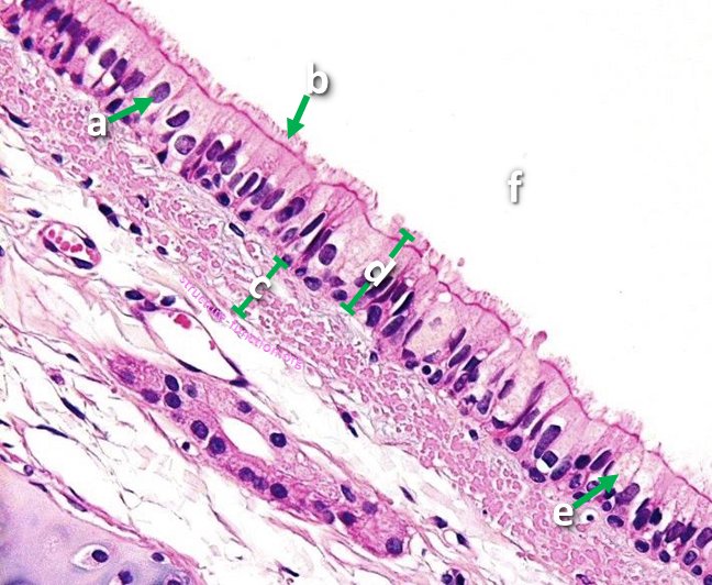
- Identify the structure indicated
- Identify the structure indicated
- Identify the structure indicated
- Classify the tissue type indicated
- Classify the cell type indicated
- Identify the structure indicated
- Provide one function for this tissue
- Provide one location for this tissue